Newest
Topics:
For the latest news, see the NEWEST TOPICS page.
Google is too dumb to let me put the list of news in this column and falsely claims that all my pages are self-duplicates.
Google-NONSENSE
Google's so-called "Artificial Intelligence" is an abuse of the concept of intelligence!
Do "IOCG" deposits form from fluids containing
abundant CO2?
(IOCG deposits = Iron Oxide Copper Gold type
deposits)
Kingsley Burlinson, June 2016, April 2018,
April 2020
Introduction
It has been asserted by Professor Murray Hitzman (SEG
international exchange lecture, 2016) and others that the fluids
from which IOCG deposits form are CO2 rich. Is this
correct? How can we determine the fluid compositions involved in
the deposition of these opaque Fe-oxide minerals? The
determination of formation fluid composition is done by using
fluid inclusions and this is almost always done by
micro-thermometry, which requires transparent minerals. It
is not possible to examine fluid inclusions in the opaque iron
oxide minerals using microthermometry. (Except for limited
studies of some haematite using infrared light.)
Almost all studies of IOCG deposit fluids are done solely on
quartz but in doing so it is assumed that the quartz and Fe-oxides
are contemporaneous and formed from a single parent fluid. However
many deposit studies show that there are multiple fluid events.
Many studies even fail to carry out proper paragenetic studies to
validate the assumption of co-genesis of Fe-oxide and quartz
formation. Some studies even completely fail to mention that
they were done entirely on quartz, a serious oversight. The
assumption of a single parent fluid forming both the quartz gangue
and the Fe-oxide minerals is unsafe.
We should be skeptical of the frequent assertions of CO2
rich formation fluids as this is almost always based upon
observation of fluid inclusions within quartz. To understand
Fe-oxide deposits we need to study the fluids in the opaque
Fe-oxide minerals. This can be done using
baro-acoustic decrepitation, infrared micro-thermometry of
some haematite samples (examples below) or by gas
extraction into a mass spectrometer during crushing or
thermal decrepitation of Fe-oxide materials.
Baro-acoustic decrepitation of numerous magnetite and haemaite
samples from IOCG deposits tentatively suggests that CO2
is not present in the parent fluids.
Assertions that IOCG deposits form from CO2 bearing
fluids
Oreskes & Einaudi, 1992, reported that a few samples
within hydrothermal quartz contained some CO2. They
found more CO2 in a few inclusions within fluorite.
Most inclusions observed did not show evidence of CO2.
They calculated that when present, the CO2 was
about 0.6 mole % (XCO2 0.006).
Bastrakov et al., 2007, studied fluid inclusions in the rare
quartz at the Olympic Dam deposit, South Australia. They used
laser raman analyses to detect CO2 in vapour
rich inclusions in either quartz or calcite (host mineral not
clearly specified) and reported that they could not detect CO2
as it was less than the detection limit.
Pollard, 2001, discussed un-mixing of CO2 bearing
fluids as a mechanism to form IOCG deposits in the Cloncurry,
Queensland. He refers to a few studies from other papers which
observed CO2 bearing inclusions in iron oxide type
deposits. All of these studies refer to fluids within quartz
only and no new observations of fluid inclusions were carried
out by Pollard.
Morales Ruano et al., 2002, studied inclusions within quartz
from the Cu deposits at Moonta, South Australia. They found no
significant CO2 in the fluid inclusions. This
deposit is hardly an IOCG type as there is no magnetite or
haematite in the late stage Cu-Au mineralized quartz veins.
None of these discussions provide significant evidence of
the involvement of CO2 rich fluids in IOCG
deposits. Only inclusions within quartz, calcite or fluorite
were studies and in some cases the conclusion was that CO2
was in fact absent. These studies are at best inconclusive in
ascertaining the association of CO2 fluids with the
iron-oxide deposition in IOCG deposits.
The above references are listed in References
at the end of this page
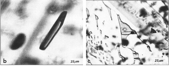
Fluid inclusion microthermometry in haematite using near
infrared illumination
Some haematite is transparent to near infrared light and can be
used for microthermometric fluid inclusion studies. But remarkably
few studies have been reported in the literature. Luders et. al
found that some haematite-quartz veins which carry gold in Brazil
do show the presence of CO2 in inclusions within
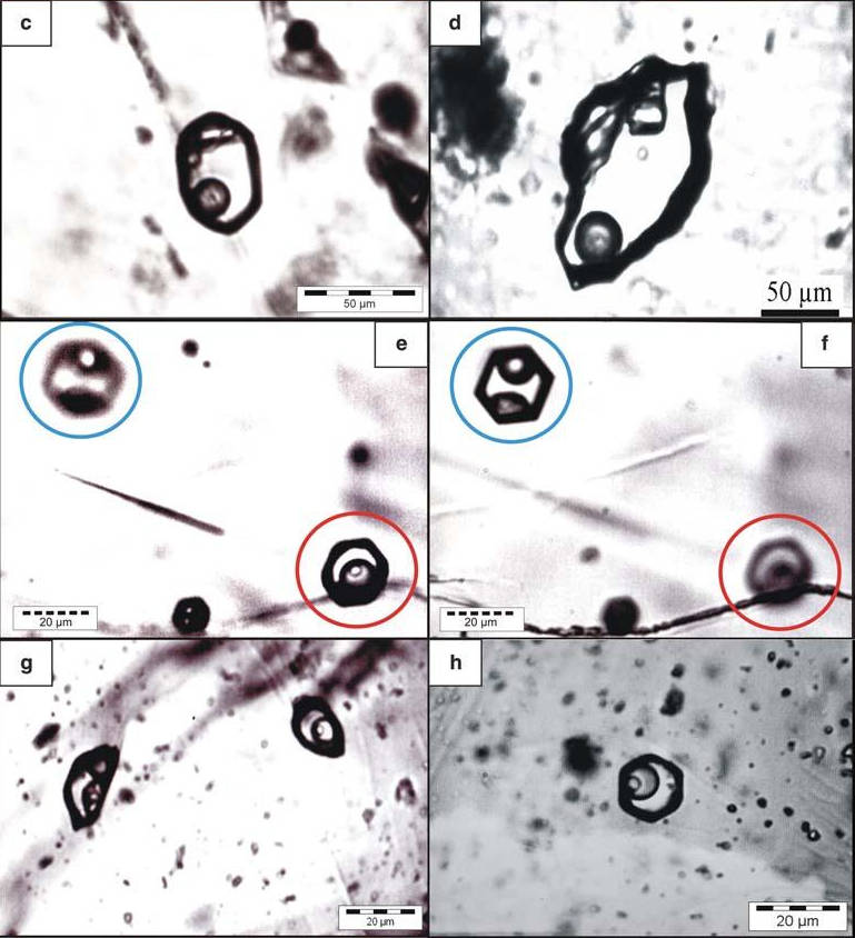
specular haematite, seen here in sub-images e, g and h.
Transmitted IR light microphotographs of fluid inclusions in
specular hematite. FROM: Genesis of itabirite-hosted Au–Pd–Pt-bearing
hematite-(quartz) veins, ́Quadrilatero Ferrıfero, Minas Gerais,
Brazil: constraints from fluid inclusion infrared
microthermometry, bulk crush-leach analysis and U–Pb
systematics. BY: Volker Luders, Rolf L. Romer,
Alexandre R. Cabral, Christian Schmidt, David A. Banks &
Jens Schneider
Mineralium Deposita (2005) 40:289 Fig. 3
Fig.3 c–h: Transmitted IR light microphotographs of fluid
inclusions in specular hematite.
c Aqueous fluid inclusion with
solids of unknown composition in specular hematite from a barren
vein in the low-strain domain of the QF, Fabrica.
d Aqueous fluid inclusion with a
solid of unknown composition in specular hematite from a
jacutinga-style vein, Gongo Soco.
e,f) Negative crystal-shaped aqueous fluid
inclusion with a solid (blue circle in e and f) and
aqueous-carbonic inclusion (red circle in e and f) in a specular
hematite grain, Itabira.
g Aqueous fluid inclusion with
solids and aqueous-carbonic inclusion in specular hematite,
Itabira.
h Aqueous-carbonic inclusion
with a small solid from a cluster of multi phase aqueous and
aqueous-carbonic inclusions.
NOTE the small size of most inclusions, usually only 5 microns
Other studies of inclusions within haematite do not show the
presence of CO2.
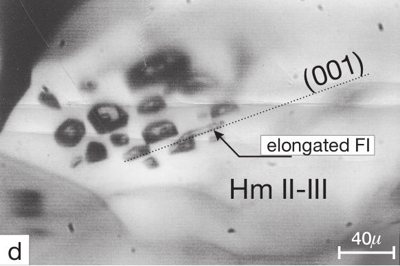
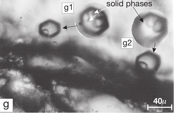
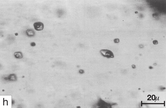
The next 3 images are From: The origin of hematite in high-grade iron ores based on
infrared microscopy and fluid inclusion studies: the example of the Conceição
mine, Quadrilátero Ferrífero, Brazil BY: Carlos Alberto Rosière & Francisco Javier
Rios
Economic Geology, (2004) Vol. 99, pp. 611–624. Fig 4
Primary two-phase fluid inclusions typical of Hm II crystals,
enclosed in an Hm II-III grain. Some of the inclusions are
elongated parallel to the basal plane and decrepitated at 345°to
350°C.
Large fluid inclusions with hexagonal shape in specular haematite.
The fluid inclusions in the left-hand side contain a small solid
saturation phase. Insets g1 and g2 are enlargements showing solid
inclusions that formed after heating. In g1 two solid phases formed
after heating to 400°C and subsequent cooling. In g2 a single solid
phase formed after heating and cooling.
Primary aqueous carbonic fluid inclusions in quartz at 25°C. Tm(ice)
= 16.6°C and Th(total) = 149° C.
The authors state that: "The quartz veins from the analyzed samples
cut across the metamorphic schistosity (S1) or interfinger with the
banded microstructure of the hematite ores. They envelop all the
early minerals, including specularite plates and are the product of
late, aqueous carbonic hydrothermal fluids of low salinity (less
than 8 wt % NaCl equiv), with total homogenization
temperatures of the fluid inclusions of approximately 330°C. These
fluids are of uncertain age and origin and did not participate in
oxidation of magnetite or Fe mineralization processes." The CO2
rich fluids seen in the quartz are apparently a late stage post
Fe-oxide event.
This pair of images are From: Fluid inclusion studies in cogenetic hematite, hausmannite, and
gangue minerals from high-grade manganese ores in the Kalahari
manganese field, South Africa. BY: Volker Luders, Jens Gutzmer & Nicolas J.
Beukes.
Economic Geology Vol.94, 1999, pp.589-596, Fig. 3
Near IR microphotographs of haematite from the Wessels mine
(Kalahari manganese field, South Africa)
b) Primary fluid inclusions decorating
growth zones in hematite.
c) Necking-down of fluid inclusions in
hematite.
Again, the inclusions lack evidence of CO2 in the
fluids.
The few studies of inclusions within haematite using infra-red
microscopy do confirm that some fluids are CO2 rich,
but in other cases the fluids lack CO2 and there are
too few studies to draw an overall conclusion about the typical
compositions of IOCG forming fluids.
Opaque mineral analysis by baro-acoustic decrepitation
Examples of decrepitation from various FeOx deposits are shown
here. Decrepitation can be intense and occurs in both haematite
and magnetite minerals.
This data shows that Fe-oxides do retain fluid inclusions and
decrepitation can provide information about formation
temperatures.
Fe-oxides generally lack the low temperature decrepitation peak
near 300 C seen in quartz containing CO2 rich fluid
inclusions. This may be interpreted as evidence that Fe-oxides do
not usually contain CO2 rich fluids. However, the
Young's modulus of magnetite (and also haematite) is much higher
than that of quartz. The increased strength of the Fe-oxide
minerals could withstand higher internal inclusion pressures
before decrepitation occurs, leading to typically higher
decrepitation temperatures than in quartz. The low-temperature
decrepitation peak caused by CO2 fluids in quartz could
be shifted to higher temperature or even be absent in Fe-oxide
minerals due to their higher Young's modulus. (A discussion of the
dependence of decrepitation upon the young's modulus of
host minerals is here.)
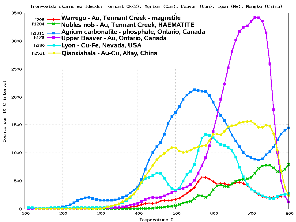
Note that the magnetite sample from the Agrium carbonatite (blue)
does show decrepitation below 300 C. This may be caused by CO2
rich fluid inclusions which would not be unexpected in a
carbonatite deposit. This suggests that decrepitation may be valid
for detecting CO2 rich fluids in magnetite and
consequently that most IOCG deposits did not form from CO2
rich fluids as they do not decrepitate below 350-400C.
Mass spectrometric analysis of gases released during sample
crushing.
The best way to be certain of the CO2 contents of
Fe-oxide minerals is by mass spectrometric analysis of the gas
released during either crushing or thermal decrepitation of
mono-mineralic haematite or magnetite.
But no such analyses have been found in the literature to
date.
Two attempts to carry out mass spectrometric analyses of
inclusion fluids have been made.
Analysis attempt 1
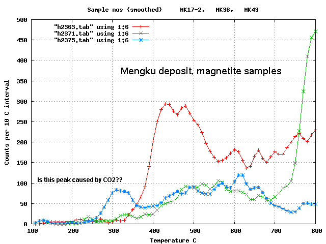
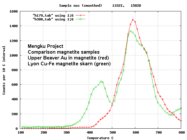
Five samples of magnetite were analysed by mass spectrometry with
this equipment by D. Gaboury to try and determine the fluid
composition. Three samples were from Mengku,
Altay area, China, one from the Lyon deposit, Nevada,
USA and one from The Upper Beaver deposit,
Ontario, Canada. The samples were chosen to be monomineralic
magnetite with significant baro-acoustic decrepitation. The
decrepitation results of the samples submitted for mass
spectrometer analysis are shown in the 2 graphs below.
No actual data files from the analyses of these samples are
available. D. Gaboury reported merely that "In short, in all 5
samples, there is no gas release related to fluid inclusion
decrepitation." This comment seems strange as it implies
there was not even water present in the gases released from these
samples. The equipment used by D. Gaboury does detect water
release, as seen here
in quartz samples. This instrument uses thermal
decrepitation of the sample to open the fluid inclusions.
There should have been some gas detected as these samples did
show significant decrepitation. The decrepitation instrument does not detect counts
caused by mineralogical effects and it is certain that fluid
inclusions are present.
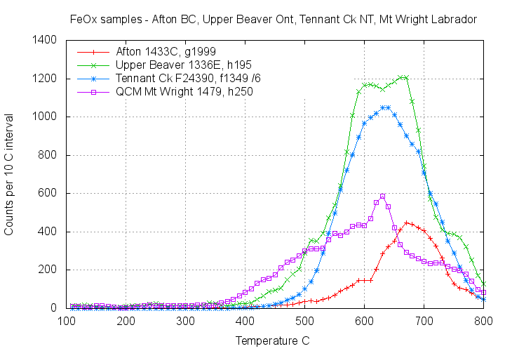
Analysis attempt 2
Another attempt to analyse the gases released from magnetite
samples by mass spectrometry was carried out by N. Blamey.
The four samples analysed were from Afton, British Columbia,
Canada, Upper Beaver, Ontario, Canada, Tennant Creek, Northern
Territory, Australia and the Mt. Wright iron mine, Labrador,
Canada. The samples were again selected to be monomineralic
magnetite with substantial decrepitation.
Decrepitation results of the 4 samples submitted for the second
attempt of mass spectroscopic examination of fluid inclusion
contents.
No actual data files of the analyses of these samples are
available. N. Blamey reported merely that "Only one of the samples
gave me gas; the material is so fine that it would appear that the
inclusions have been compromised". Neither the gas found nor the
sample it came from were identified. The samples provided were
part of the same material that was decrepitated and was sieved to
<420 and >200 microns. It was not so fine grained that the
fluid inclusions were compromised as can be seen from the above
decrepitation graphs of these samples.
Why did the mass spectrometric analyses fail to detect any
inclusion fluids?
It is clear from the decrepitation results that there are
inclusions in the magnetite samples, although perhaps less
numerous than in typical quartz samples. And in all cases, the
sample material decrepitated and analysed by mass spectrometry was
identical. Decrepitation counts are not due to crystallographic or
mechanical effects and repeat analyses of
already analysed samples give no counts. Only irreversible
events are counted, so it is certain that decrepitation counts are
caused by fluid inclusions.
Perhaps the inclusions in magnetite are so small that they do not
release enough gas to be measured in the mass spectrometer. In the
photographs of FIs in haematite (above)
most inclusions are very small, often just 5 microns across.
Decrepitation of such small inclusions would give very little gas
to analyze. In the mass spectrometer, the flux of analyte into the
ioniser must exceed the rate of evacuation by the vacuum system. A
small gas quantity released from small inclusions may well be
evacuated so fast that it is not detected in the mass
spectrometer. Typically, spectrometers require more than a
milligram of sample.
It seems that mass spectrometer analyses of fluids released by
thermal decrepitation on magnetite must be done on much larger
samples than normal to compensate for the apparently small size
and low abundance of inclusions in magnetite and haematite. In
both these studies the analyst did not provide details on the
sample size they used.
Conclusions
There have been very few FI studies of haematite by infrared
microthermometry. CO2 rich fluids have been seen
in one study, but in others the haematite lacks CO2
while adjacent quartz is CO2 rich, indicating different
fluid events.
There is only minor evidence for the presence of CO2
bearing fluids in these deposits and all of that is based on fluid
inclusion studies in quartz or other gangue minerals and is at
best very questionable evidence for the presence of CO2
fluids in the formation of the Fe-oxide and economically
interesting minerals of concern.
Most fluid information on IOCG deposits is actually derived from
FIs within quartz. Often there is no paragentic study and it is
uncertain that the quartz and Fe-oxides are actually deposited
from the same fluid.
Baro acoustic decrepitation of haematite and magnetite almost
always lacks the low temperature decrepitation
peak caused by CO2 rich fluid inclusions hosted in
quartz suggesting that CO2 rich fluids are not
involved. But the young's modulus of both magnetite and haematite
is about double that of quartz, so it is not conclusive that CO2
fluids within Fe-oxides would cause the same characteristic
low-temperature decrepitation peak as seen in quartz.
No mass spectroscopic analyses of gases extracted during crushing
or thermal decrepitation of Fe-oxides have been found in the
literature. Attempts to analyze gas extracted from 9 magnetite
samples by thermal decrepitation have failed to find any gas in 8
of the samples, not even water! One sample contained unidentified
gas.
The inability to measure gas extracted form these samples which
have substantial decrepitation responses is probably because
insufficient fluid was released from the fluid inclusions. It
seems that inclusions in haematite and possibly magnetite may
typically be smaller than inclusions in quartz, hence containing
less fluid. Baro-acoustic decrepitation of magnetite samples also
is usually less intense than quartz samples, suggesting fewer
inclusions. These effects could reduce the fluid amount to less
than is required to perform the mass spectrometric analysis. Much
larger samples may be needed to analyse magnetite fluids. As the
analysts involved have not provided the actual data files it is
not possible to be certain of this explanation and further more
careful mass spectroscopic work with larger samples is required.
Recent studies using stable isotopes of Cu (Saunders et al,
Mineralium Deposita 2016, V51 #1) have confirmed different fluid
sources for ore and gangue minerals in epithermal Au-Ag deposits.
The authors state: "This conclusion has implications for fluid
inclusion and isotope studies that have focused on using the
gangue minerals for analysis, if those minerals do indeed have
principally different sources." This is a serious concern for
Fe-oxide deposits as FI studies are almost always done only on the
quartz gangue minerals.
There is no clear evidence that CO2 bearing fluids
are coeval with or involved in the deposition of Fe-oxide
minerals and the associated Cu and Au minerals.
========================================
References
Bastrakov EN, Skirrow RG, Davidson GJ (2007) Fluid
evolution and origins of iron oxide Cu-Au prospects in the Olympic
Dam District, Gawler Craton, South Australia. Econ. Geol.
102:1415-1440.
Morales Ruano S, Both RA, Golding SD (2002) A fluid inclusion and
stable isotope study of the Moonta copper-gold deposits, South
Australia: evidence for fluid immiscibility in a magmatic
hydrothermal system. Chem. Geol. 192:211-226
Oreskes N, Einaudi MT (1992) Origin of hydrothermal fluids at
Olympic Dam:preliminary results from fluid inclusions and stable
isotopes. Econ. Geol. 87:64-90
Pollard PJ (2001) Sodic (-calcic) alteration in Fe-oxide-Cu-Au
districts: an origin via un-mixing of magmatic H2O-CO2-NaCl
± CaCl2-KCl fluids. Mineralium Deposita 36:93-100
Saunders, J.A., Mathur, R., Kamenov, G.D., Shimizu, T. and
Brueseke, M.E. (2016), New isotopic evidence bearing on
bonanza (Au-Ag) epithermal ore forming processes. Mineralium
deposita 51:1 p1
========================================
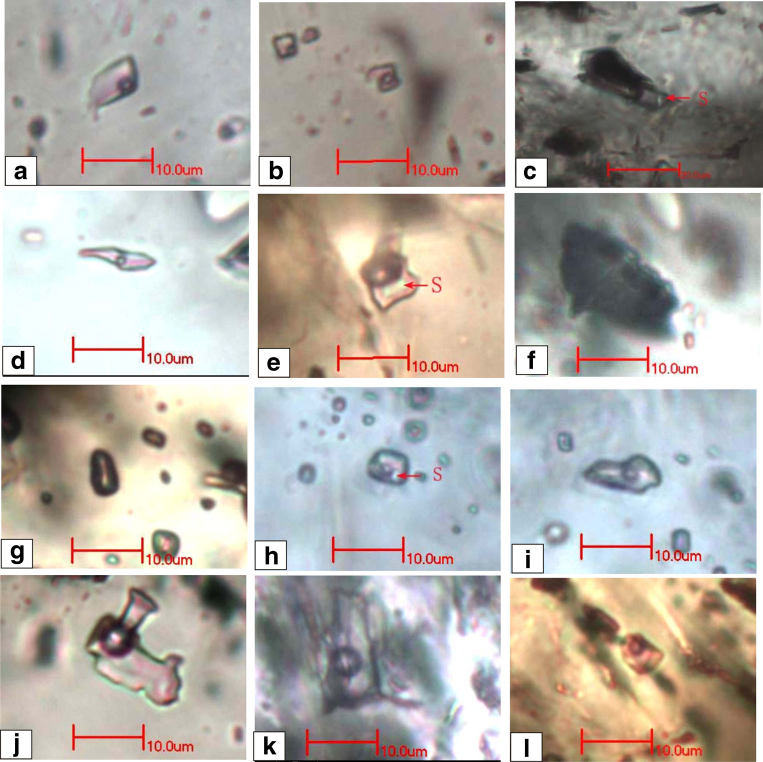
Appendix - additional FI photographs in iron oxide associated
minerals.
Fluid inclusion images in apatite, siderite, quartz and carbonates
of the Meishan feox-apatite deposit. (Fluids for Fe and late Au are
different!)
FROM: Mineralium deposita 50:7 p847 2015 Yu et. al.
d,e,f & j are within quartz. a,b & c are
within apatite. j,h &i are within
siderite k is in calcite
l is in dolomite NOTE the small size of most inclusions, usually only 5 microns
Author's figure caption: a,b two-phase inclusion in stage 2 apatite, c three-phase inclusion (L+V+S) in stage 2 apatite, d two-phase inclusion in quartz as cavity fillings, e three-phase inclusion in quartz as cavity fillings, f vapor-phase inclusion in quartz as cavity fillings, g vapor-phase inclusion in siderite, h three-phase inclusion in siderite, i two-phase inclusion in siderite, j two-phase inclusion in quartz as vein, k two-phase inclusion in calcite as vein, l two-phase inclusion in dolomite as vein
S=solid phase
 Applied Mineral Exploration