A few samples from the Krivoy Rog Iron deposits
in Ukraine have been studied to compare them with other
Banded
Iron Formation (BIF) type deposits in an ongoing project
in association with
Marta
Sosnicka of AGH University, Krakow, Poland.
Krivoy Rog is in southern Ukraine some 200 Km from the Black Sea
coast.
 A
technical article discussing the Krivoy Rog deposits is
available in Economc Geology, Nov 1973.
A
technical article discussing the Krivoy Rog deposits is
available in Economc Geology, Nov 1973.
Krivoy Rog is within the
Kursk
magnetic anomaly, one of the largest and most intense on
earth. The Krivoy Rog area is a major source of Fe with many
active mines.
An initial suite of 12 samples was studied, with 3 additional
samples provided later.
The sample
details are here.
Magnetite samples were obtained from the Frunze underground
mine, along with a sample of martite.
For reference, quartz samples were obtained from 3 nearby areas
of surface outcrop. And 3 additional samples, K1, INK11 and V3
from the Yugok pit were also studied later.
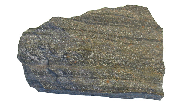
The iron deposit is a metamorphosed Banded Iron Formation and
this photo is a museum specimen sample from Krivoy Rog,
photographed by Siim Sepp.
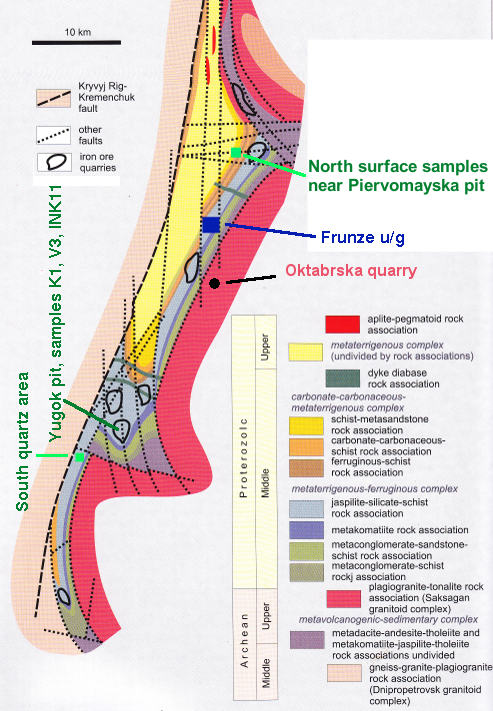
The sample locations are shown on this map, which is from:
Bobrov, O.B., Gurskiy, D.S., Krasnozhon, M.D., Malyuk, B.I.,
Shcherbak, M.P., Kalinin, V.I., et al., 2002, Main types of
rock complexes and mineral deposits in the Ukrainian Shield,
Geological Excursion Guidebook, Ukrainian State Geological
Survey, Kiev.
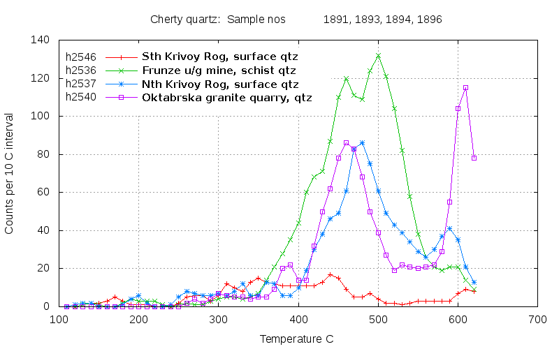
During sample preparation, the hand specimens were described.
Some of the quartz samples were described as cherty because they
were very fine grained and of vitreous appearance, while others
were considered to be of hydrothermal quartz vein origin as they
were coarsely crystalline. When decrepitated, these two types of
quartz are quite distinct as chert gives no significant
decrepitation because it formed at low temperatures and fluid
inclusions are either very small or absent. Samples 1892, 1893
and 1894 were described as being of cherty appearance and these
show very weak or no decrepitation here. These samples may be
comprised dominantly of sedimentary banded chert silica. In
addition, sample 1894 (analysis h2540) also has weak
decrepitation, although it was not described as being cherty and
cannot be a chert as it crosscuts the granite!
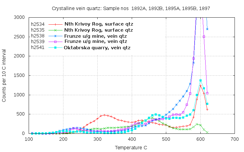
The vein quartz samples show typically intense decrepitation
confirming a hydrothermal or metamorphic origin. 3 of these 5
samples show low temperature decrepitation caused by CO
2
rich fluid inclusions. But note that sample 1892A (analysis
h2534, red) is very different from sample 1892B (analysis h2535,
green) despite being collected close together. The 2 quartz
samples from the Frunze mine, shown in blue and magenta (h2538
& h2539) are similar and contain some CO
2 rich
fluid inclusions which decrepitate at 230 C, which is a
much lower temperature than the surface quartz samples. This low
temperature may be caused by a very high partial pressure of CO
2
in the inclusions from the Frunze mine quartz.
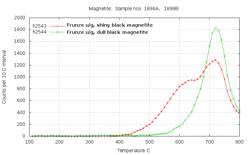
The magnetite samples from the Frunze mine show considerable
decrepitation, not unlike magnetites from skarn deposits. Sample
1898A (red) has a prominent double peak indicating 2 very
distinct fluid inclusion populations are present. The inclusions
in these magnetites are thought to have been introduced during a
regional metamorphic event as these deposits are not considered
to be skarns of high temperature hydrothermal origin.
Unmetamorphosed sedimentary magnetites would not decrepitate at
all as they would lack the necessary high pressure/temperature
fluid inclusions.
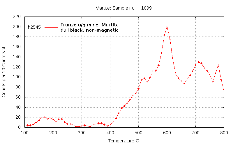
Martite is an alteration mineral, a pseudomorph of haematite
after magnetite. It shows only a minor amount of decrepitation
because it lacks fluid inclusions, which were probably destroyed
during the alteration of the parent magnetite.
The second group of samples, V3 (2137), K1
(2139) and INK11 (2138), from the Yugok open pit.
The second group of samples included number V3 which was
split into 2 subsamples, 2137A (crosscutting quartz) and 2137B
(host BIF) during preparation.
These samples were further split into magnetic and non-magnetic
fractions and also coarse grainsize (<420, >200 micron)
and fine grainsize (<200, >100 micron) fractions before
being analysed.
The different aspects of these 3 samples can be seen in this
photo of sample 2137 (V3) including crosscutting clean quartz
vein material (2137A) and the fine grained banded chert -
magnetite BIF (2137B).
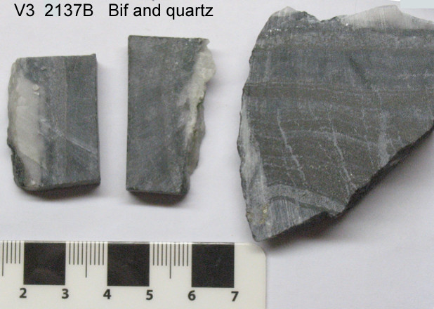
Sample 2137 - V3 (Vein 3 in the paper by M. Sosnicka
et. al.)
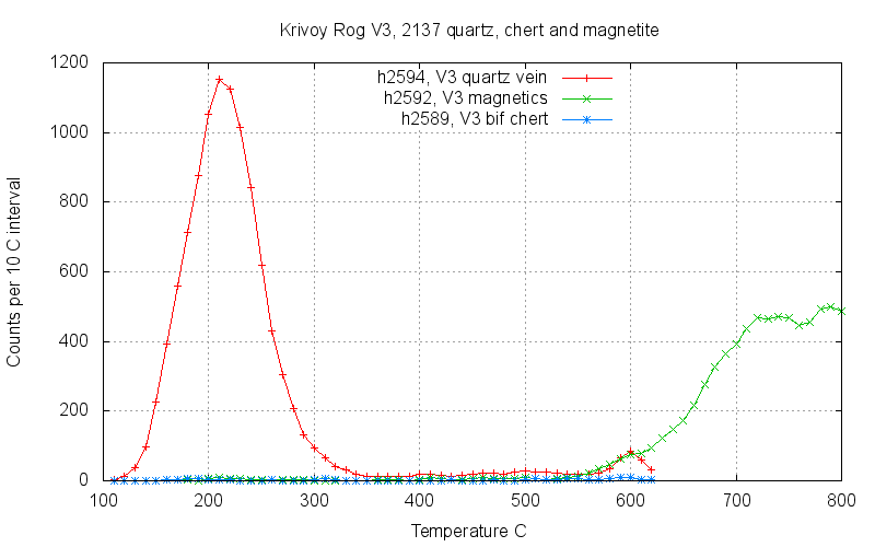
The crosscutting quartz in sample 2137B , shown here in red, had
extremely intense low temperature decrepitation near 200 C. Low
temperature decrepitation such as this is usually caused by high
CO
2 contents in the inclusions. However this peak is
more intense and at lower temperature than is usual.
Crushed grains of the sample were immersed in a refractive index
oil (clove oil) to try and identify the cause of this unusual
decrepitation behaviour. Abundant fluid inclusions were seen,
but they were only small, typically 5 to 8 microns across only.
And the fluid content was usually dominated by a single vapour
phase with very little water present. Angular inclusion
contents were very common indicating the presence of a solid
phase. This was not cubic and could not be halite, and was
unidentified in these observations. Bubbles were seen only
rarely in the smaller inclusions which were less than 2 microns
across. These observations failed to identify a CO
2
rich fluid which could cause the decrepitation. In the other
samples, 2138 and 2139,
M. Sosnicka
identified abundant nahcolite ( NaHCO
3 ) solid phases
in the inclusions.The unidentified solid in sample 2137 is
probably also nahcolite. Nahcolite decomposes at 270 C giving
off CO
2 (at 1 atmosphere pressure) and it seems
likely that the low temperature decrepitation peak is caused by
decomposition of nahcolite in combination with already high CO
2
contents in the inclusions.
The magnetic fraction from the BIF (shown in green) in this
sample (2137A) does not decrepitate at low temperature,
presumably because the magnetite does not contain nahcolite.
Decrepitation in the magnetite occurs only above 600 C and is
interpreted to be the result of inclusion growth during
recrystallization at the known regional metamorphic conditions.
The non-magnetic chert fraction from the BIF (shown in blue) in
this sample (2137A) does not decrepitate at all, which is
typical of fine grained cherts, regardless of subsequent
metamorphism.
It is clear that the fluids within the crosscutting quartz vein,
2137B, are quite different to those in the BIF magnetics, 2137A.
Although this is not really surprising, it does highlight the
necessity to avoid assuming that fluid inclusions in quartz are
representative of the formation conditions of associated opaque
minerals. This is also discussed for pyrite samples
here.
Quartz is always used to study fluid inclusions simply because
it is transparent, a requirement for microthermometry. But this
data from quartz is then assumed to apply to co-existing opaque
minerals. This decrepitation data shows that this assumption is
frequently invalid.
The metamorphic fluids which upgraded the BIF to ore grade
magnetite removed silica. But the fluids in the crosscutting
quartz vein were clearly depositing silica and cannot tell us
much about the fluid environment during ore formation.
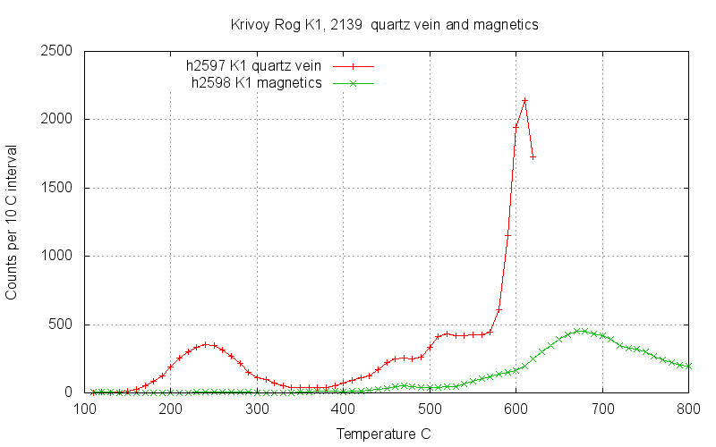
Sample 2139 also shows a prominent low temperature decrepitation
peak in the crosscutting quartz vein, shown in red. This is
again presumed to be due to the presence of nahcolite which M.
Sosnicka identified in this sample. The peak is at slightly
higher temperature but the reason for this is unknown. But there
are also other populations of fluid inclusions in this sample
which did not occur in sample 2137.The decrepitation in quartz
(red curve) at 460 C is matched with decrepitation in magnetite
(green curve). This and the higher temperature decrepitation
peaks in the quartz may well represent the same fluids that
cause the decrepitation peak at 670 C in the magnetite. The
chert from the BIF was not analysed.
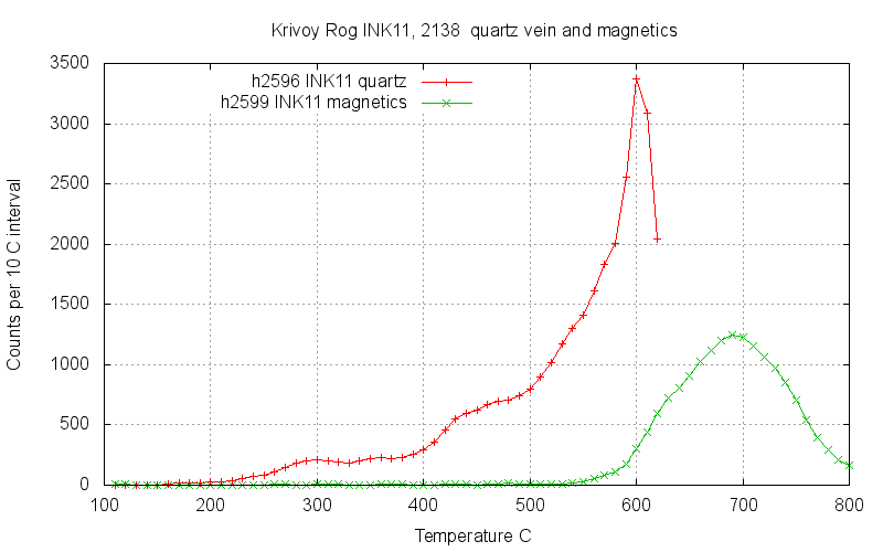
Sample 2138 shows only a weak low temperature decrepitation and
at much higher temperature than in sample 2139. Although
nahcolite was identified in this sample by M.Sosnicka, it is not
clear why the intensity of decrepitation is so much lower or why
the temperature so much higher. The chert from the BIF was not
analysed
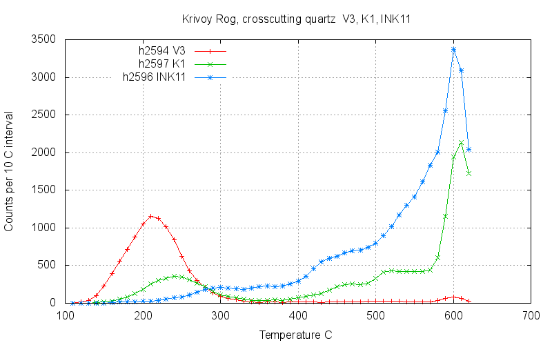
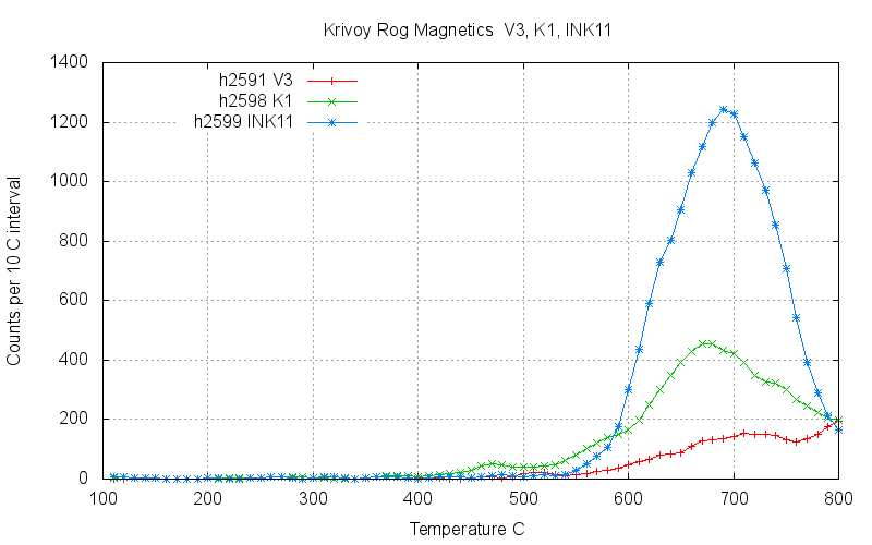
 The following graphs show the same results as above but
grouped by sample type instead of sample number to highlight
the similarities and differences within this suite of samples.
The following graphs show the same results as above but
grouped by sample type instead of sample number to highlight
the similarities and differences within this suite of samples.
The crosscutting quartz samples shows the change in temperature
and intensity of the low temperature decrepitation. There is not
enough data to attribute this change to variations in the amount
of nahcolite present, although this may be the cause.
The magnetic fractions from the BIF show similar high
temperature decrepitation on all of the samples, as would be
expected with inclusions resulting from a regional metamorphic
event. However, there are also some interesting differences and
further studies are required to understand these changes.
 Sample 2137 was also used for
comparisons between different analytical grainsize fractions
and magnetic fractions and this information is discussed here.
Sample 2137 was also used for
comparisons between different analytical grainsize fractions
and magnetic fractions and this information is discussed here.
There are also a number of decrepitation studies of magnetites
and haematite reported elsewhere on this website.
Great Bear magmatic zone, Canada.
Mengku,
China.
Tennant
Creek, Australia.
An
overview of FeOx data. and
additional
FeOx data.
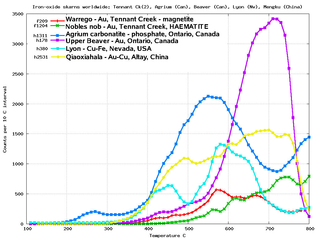
For comparison, here are some typical decrepitation results from
other magnetite deposits and skarns.
 An additional discussion contrasting
Skarn magnetites with BIF magnetites is here.
An additional discussion contrasting
Skarn magnetites with BIF magnetites is here.
Summary
Decrepitation is an effective way to study magnetites and
haematites because you cannot use optical microscopy on these
minerals, leaving very few other methods to obtain fluid
inclusion data. Although fluid inclusions within these opaque
minerals are less well understood than inclusions within
transparent minerals, the same principles apply and we can see
marked differences between deposit styles and samples. The
method can be used to help discriminate between low temperature
and hydrothermal origins for Fe-oxide minerals. For example, it
distinguishes between skarn and BIF deposits.
Although CO
2 rich fluid inclusions within magnetite
should produce the same low temperature decrepitation effect
that we
frequently see on
quartz samples, very few magnetites have such low
temperature decrepitation peaks. The 3 magnetite samples from
the Frunze mine and Yugok pit at Krivoy Rog do not show low
temperature decrepitation and it is inferred that the fluids
here were aqueous and had very low CO
2 content. Such
low CO
2 content fluids seem to be a feature of
magnetite samples from BIF deposits.
However there are two quartz samples in the Frunze mine which do
have a (small) low temperature CO
2 decrepitation peak
(1895A & 1895B; H2538 & H2539) as well as an intense CO
2
decrepitation peak on quartz from the Yugok pit (2137; H2594).
This suggests that there were multiple fluid stages.
Although many more samples are required to understand this
system, perhaps there was an early stage pervasive event
(regional metamorphic?) with low CO
2 content fluids,
and subsequent quartz veining with a different and CO
2
rich fluid with only localised influence.
The microthermometric studies by M. Sosnicka show that the
crosscutting quartz samples were formed from a CO
2
rich fluid which also deposited nahcolite. This fluid was
determined to have come from a nearby carbonate-containing rocks
during the regional metamorphism.
Acknowledgement: Special thanks to M. Sosnicka for
providing these samples and for associated geological
information.
References
Fluids in the metamorphic stage of Krivoy Rog iron deposit
evolution, Ukraine. Sośnicka,
Marta, Bakker, Ronald J. European
Current Research on Fluid Inclusions (ECROFI-XXI),
Leoben, Austria, 2011. Abstracts, p. 182
Fluid types and their genetic meaning for the BIF-hosted iron
ores, Krivoy Rog, Ukraine.
Marta Sośnicka, Ronald J. Bakker,
Curt Broman, Iain Pitcairn, Ihor Paranko, Kingsley Burlinson.
Ore Geology Reviews 68 (2015) 171–194
Abstract here
Extract
(PDF file)(3.5 Mbyte) here
 Applied Mineral Exploration